HomeProjectsBiomedical Sciences and Healthcare TechnologiesNovel Imaging Approach Brings Faster 3D Imaging for Biomedical Researches

Novel Imaging Approach Brings Faster 3D Imaging for Biomedical Researches

Project Quick Facts

Principal Investigator

  • Prof. CHEN Shih-Chi

    Department of Mechanical and Automation Engineering

  • Funding

    Research Grants Council, Innovation and Technology Commission

Activities of neurons are generally completed on a time scale of 10 milliseconds, which makes it hard for conventional microscopes to observe these phenomena directly. On the contrary, two-photon microscopy works by delivering ultrafast pulses of infrared laser light to the sample, where it interacts with fluorescent labels to create an image. It is extensively used for biological researches because of its ability to produce high-resolution 3D images up to a depth of one millimeter in a living tissue or to monitoring activities from hundreds of neurons simultaneously. These advantages, however, come with a limited imaging speed of the two-photon microscopy because of the weak fluorescent signal.

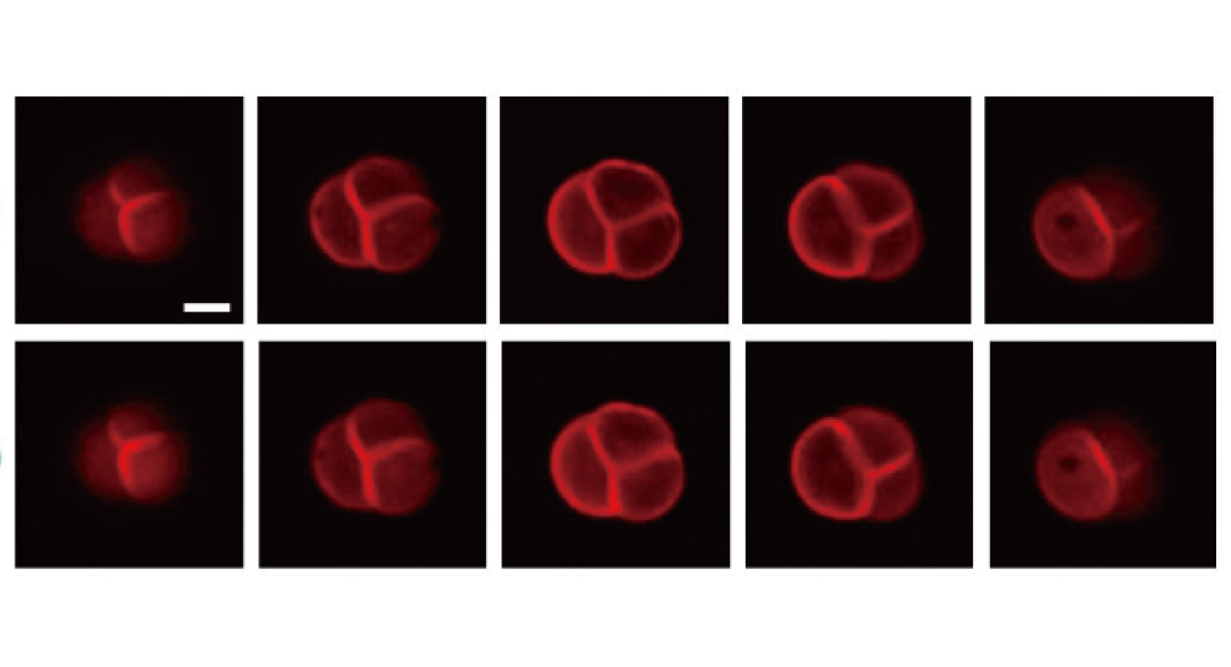
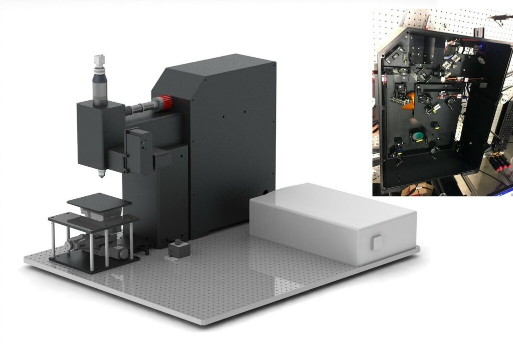
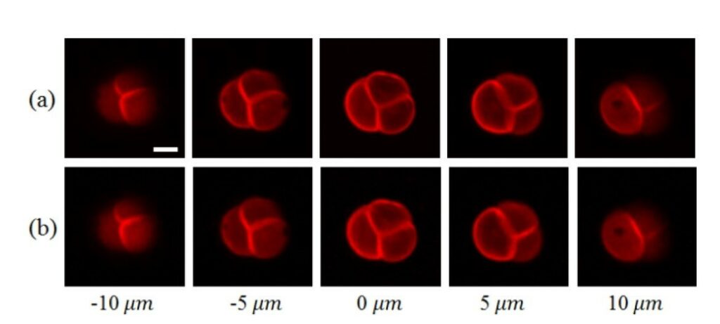
To speed up scanning, this project developed a multi-focus laser illumination method that uses a digital micromirror device (DMD). The new approach is able to produce two-photon microscopy images of a 3D sample in one second without sacrificing the resolution, which is at a speed three to five times that of the conventional point-scanning method. The research solves the problem of conventional DMD being unusable to work with ultrafast laser, enabling them to be integrated and used in beam shaping, pulse shaping, and two-photon imaging. We further increased the imaging speed in this research by combining multi-focus scanning with compressive sensing. This approach enables image acquisition with fewer measurements. This is because it carries out image measurement and compression in a single step and then uses an algorithm to rebuild the images from the measurement results. Experiments demonstrated the technique’s ability to produce high-quality 3D images with high imaging speeds from any field of view. For example, they were able to acquire 3D images from a pollen grain, in just 0.55 seconds. The same images acquired with traditional point scanning took 2.2 seconds.

Reference:

C. Wen, M. Ren, F. Feng, W. Chen, and S. Chen, “Compressive Sensing for Fast 3-D and Random-access Two-photon Microscopy,” Optics Letters, 44(17): 4343-46, 2019.
We have developed a system that increases the imaging speed of two-photon microscopy up to three to five times without compromising resolution. On the left is a CAD model of the compact, custom built, two-photon microscopy system. The free-space optics inside are pictured on the right.
We prepared two-photon microscopy images of a pollen grain by using (a) traditional point-scanning and (b) the new compressive imaging approach. The point-scanning imaging time was 2.2 seconds while the compressive imaging time required only 0.55 seconds.

Do you like our project?

MORE TO EXPLORE