HomeProjectsBiomedical Sciences and Healthcare TechnologiesClinical Evaluations and Applications of Innovative Optical Coherence Tomography Imaging System (OCTIS) in Endometrium

Clinical Evaluations and Applications of Innovative Optical Coherence Tomography Imaging System (OCTIS) in Endometrium

Project Quick Facts

Principal Investigator

  • Prof. LI Tin Chiu

    Department of Obstetrics & Gynaecology

  • Prof. WANG Chi Chiu

    Department of Obstetrics & Gynaecology

  • Funding

    Innovation and Technology Commission

  • Collaboration

    Tomophase Ltd., and
    Guangdong Winstar Medical Technology Co. Ltd.

  • Sponsorship

    Sponsorship Test

  • Patent

    China patent filed

  • Award

    Champion, Nanshan Entrepreneur Star 2018, Shenzhen, China
    Biomedical-industry Champion, Nanshan Entrepreneur Star 2018, Shenzhen, China

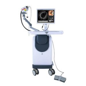
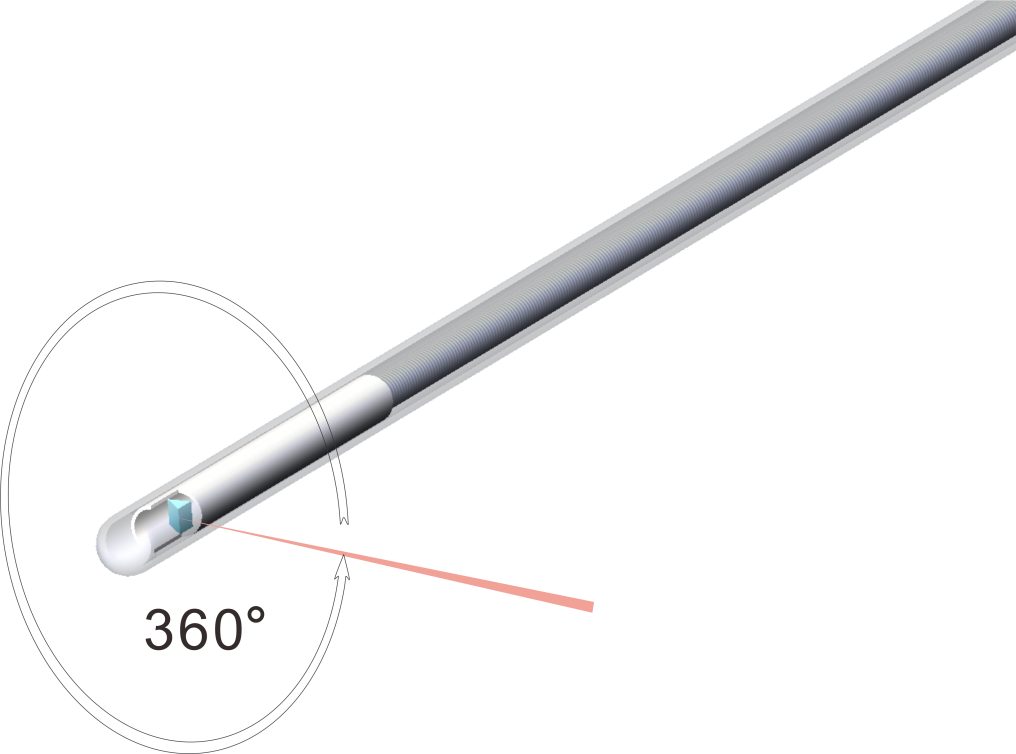
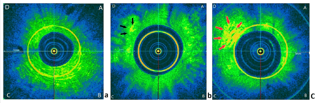
Half of the causes of women’s infertility are related to the uterus, but the existing methods for examining the endometrium are external, and the resolution of the image is low, such as ultrasound, MRI, etc. Hence, doctors failed to distinguish whether the endometrium has inflammation, injury or defects etc., which may affect the chance of conception. Department of Obstetrics & Gynaecology from CUHK adopted the first Optical Coherence Tomography Imaging System (OCTIS) for endometrium in the world. OCTIS incorporates catheter-based rotational and pullback probes with improved OCT technologies. It can be used in endoscopy for ductal organ imaging. CUHK team carried out comprehensive clinical evaluations of OCTIS in endometrium imaging for 20 women, and then tried to discover the underlying cause of the disease. As a result, OCTIS can improve the pregnancy outcome in women with reproductive failure undergoing embryo transfer.

Optical Coherence Tomography Imaging System (OCTIS)
Catheter-based rotational and pullback probe

Uniqueness and Competitive Advantages:

  • Less harmful to patients. It is because this system is minimally/non-invasive, and it uses near infrared non-ionizing radiation. Additionally, there is no side effects of hysteroscopy.
  • Better imaging. This system provides real-time imaging for pathological diagnosis. Those images are high-resolution, scanned cross-sectionally and longitudinally. Thus, we can obtain potentially microscopic structural and 3D reconstruction.
Left pic: A normal endometrium. Middle and right pics: two different diseased endometriums.

Do you like our project?

MORE TO EXPLORE

Handheld High-Resolution Short-Wave Infrared Spectrometer

Spectrometers are widely used in many different aspects, such as food and environmental inspection etc. However, it is necessary to have a portable and high-resolution spectrometer for real-time on-site inspection at remote farm or factory. CUHK team invented two new technologies, which broke the traditional boundary. We develop a handheld high-resolution short-wave infrared (SWIR) spectrometer […]

Read more >