Abstract:
Autophagy is a well-defined catabolic mechanism whereby cytoplasmic materials are engulfed into a double-membrane structure termed autophagosome. Autophagy plays an essential role in quality control of proteins and organelles, and protects the cell against pathogen infection or other unfavorable conditions. During autophagy, autophagosome engulfs and delivers cargo(s) into the lysosome/vacuole for degradation and recycling. However, little is known about the underlying mechanisms of autophagosome formation in plants.
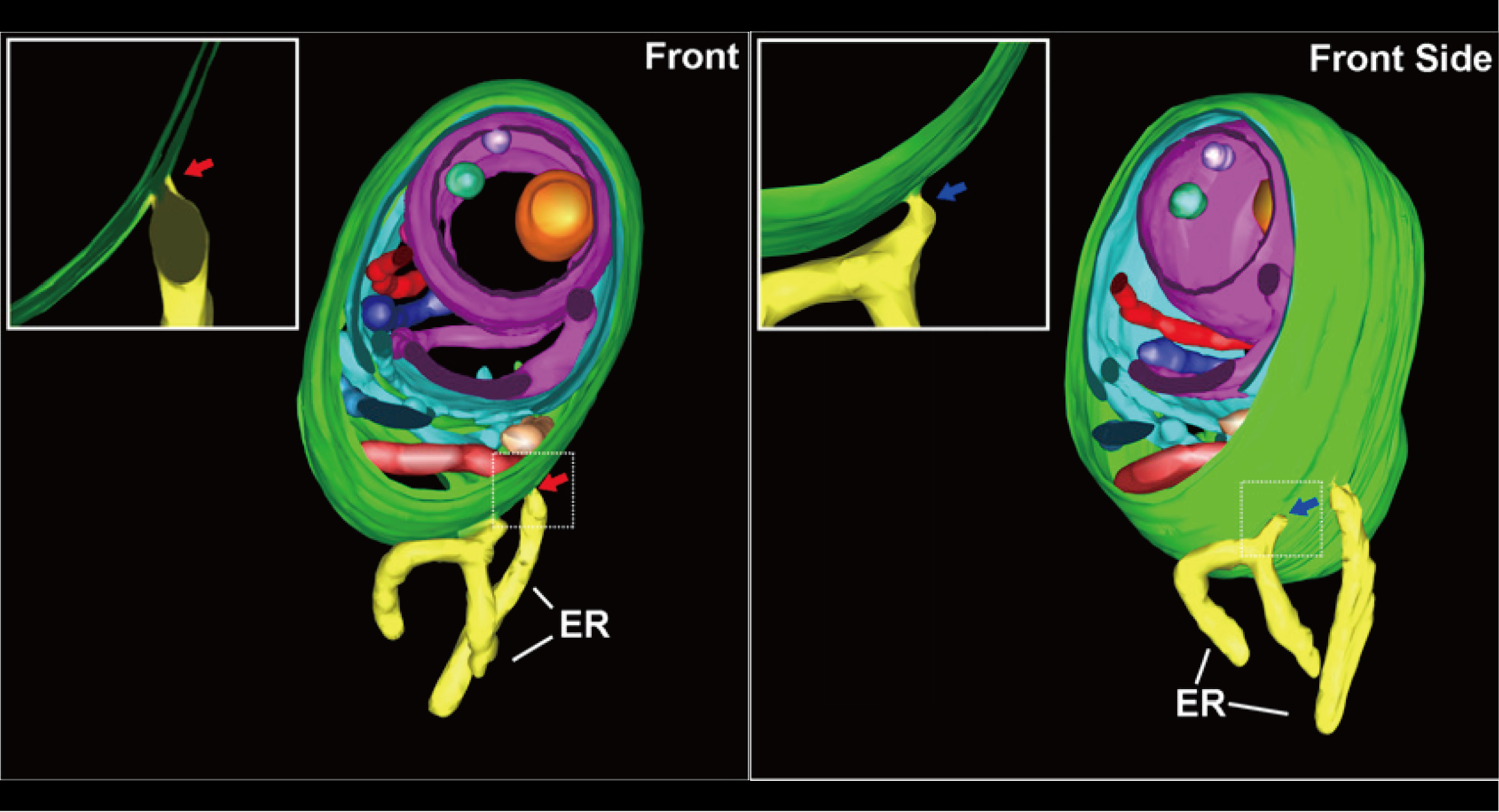
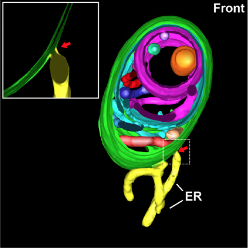
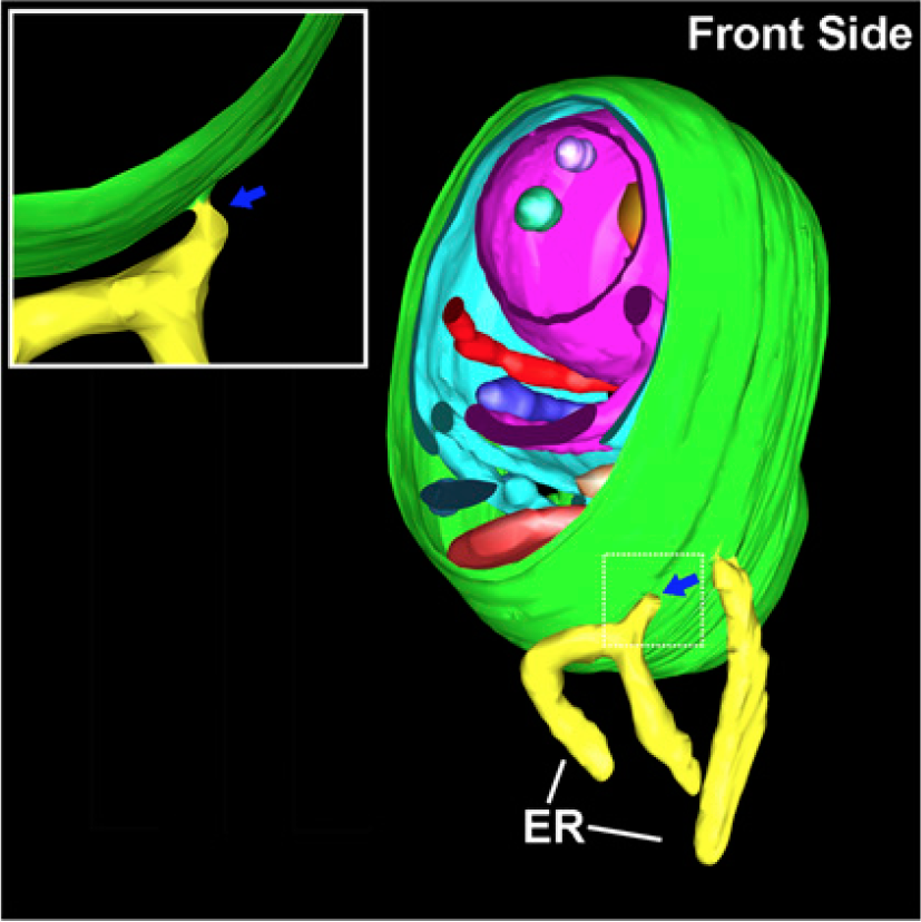
Our recent research programs have made a significant contribution towards our understanding about unconventional protein secretion (UPS)-mediated protein traffic and autophagosome biogenesis in plants. For example, using the Arabidopsis Exo70E2 (AtExo70E2) as a marker for exocytosis, we discovered a novel organelle termed EXPO (Exocyst-positive Organelle) and a new UPS unique in plants. Using AtSH3P2 and GFP-AtSH3P2 fusion as tools, we have recently illustrated the dynamics and ultrastructures of autophagosome formation in Arabidopsis as well as the essential role of AtSH3P2 in regulating autophagosome biogenesis in plants.
The goal of this project is to understand the underlying mechanisms of the E2-exocyst complex recruitment and function in plants as well as the functional relationship between EXPO and autophagosome during autophagy in plants.
Applications:
Since both EXPO and autophagosome play important roles in regulating plant growth and development as well as plant response to environment, our studies have contributed greatly towards our understanding about organelle biogenesis and function in plants, as well as their potential application in biotechnology for improving human health.